

IRON DEFICIENCY ANEMIA
1. Increased iron absorption is seen in: Iron deficiency anemia, pregnancy, hypoxia, acidic pH of stomach, ferrous iron salts
2. Causes of IDA: Hookworms, celiac sprue, carcinoma colon
3. Features diagnostic of IDA: Decreased serum ferritin, increased TIBC
4. Earliest sign of IDA: Decrease in serum ferritin
5. Most sensitive and specific test for diagnosing IDA: Serum ferritin levels
6. Hypochromia may be preceded by microcytosis
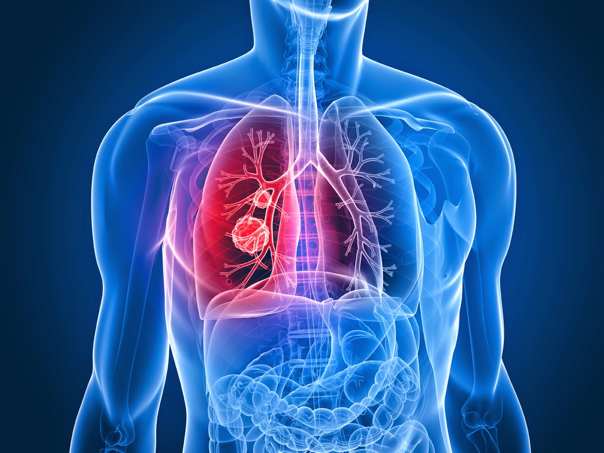
LUNG CARCINOMA
1. Most common type in non-smokers: Adeno Ca
2. Most common type in peripheral location: Adeno Ca
3. Most common type in young patient: Adeno Ca
4. Most common type in females: Adeno Ca
5. Best prognosis among lung Ca: Squamous cell Ca
6. Most common metastasizing to opposite lung: Adeno Ca
7. Lung Ca most responsive to radiotherapy: Small cellCa
8. Lung Ca most sensitive to chemotherapy: Small cell Ca
9. Most common to produce hypercalcemia: Squamous cell Ca
10. Most common cavitating lung Ca: Squamous cell Ca
11. Most common type of lung Ca: Adeno Ca
12. 2nd Most common lung Ca: Squamous cell Ca
HYPER
KALEMIA -increased level of pottasium in blood.
1. Hyperkalemia is associated with:
a. Digitalis intoxication,
b. Beta blockers,
c. Acute intravascular hemolysis,
d. Acute oliguric states
2. Severe crush injury,
3. Pseudohyperkalemia: Hemolysis of blood sample or the release of potassium from RBC, WBC and platelets
4. Serum potassium more than 5.5 mEq/ L,
RESPIRATORY FAILURE TYPES
1. Type I:
a. Parenchymal disease,
b. ARDS,
c. Pneumoniae,
d. Emphysema
2. Type II:
a. COPD,
CLINICALLY IMPORTANT SPOTS
1. Koplik spots: Measles
2. Roth spots: SABE
3. Bitot's spots: Vitamin A deficiency
4. Herald spot: Pityriasis rosea
BRONCHIECTASIS
1. Nodular bronchiectasis is seen in infection with: Mycobacterium avium
2. Chest x-ray shows: Tram track lines
3. Most common site: Left lower lobe
4. Clubbing is seen
5. Investigation of choice: HRCT scan
ASTHMA
1. Constriction of small airways,
2. Curschmann's spirals seen,
3. Increased leukotrienes,
4. Hyper-responsive airways,
5. FEV1 improves maximum with bronchodilator therapy
EMPHYSEMA and CHRONIC BRONCHITIS
1. Breathlessness is a characteristic feature,
2. Alphal antitrypsin deficiency is associated with: Panacinar emphysema
3. Smoking is an important predisposing factor,
4. Diurinal variation in peak expiratory flow rate is seen
KARTAGENER SYNDROME
1. Situs inversus,
2. Bronchiectasis
3. sinusitis
PLEURAL EFFUSION
1. Increased amylase in pleural fluid:
a. Pancreatitis,
b. Esophageal perforation
c. Burns,
d. Adrenal insufficiency
2. Blood stained pleural effusion:
a. Pulmonary infarction,
b. Metastatic Ca etc.
3. Low glucose in pleural fluid:
a. RA,
b. Empyema
4. Exudative pleural effusion is seen in:
a. RA,
b. Bronchogenic Ca etc.
5. ECG changes:
a. Shortening of QT interval,
b. PR interval prolongation,
c. Tall, peaked T wave
METABOLIC DISORDERS
1. Metabolic alkalosis:
a. Raised HCO3-,
b. CO2 unchanged (raised pH)
2. Respiratory acidosis:
a. Raised CO2 and
b. Raised HCO3- (reduced pH)
3. Metabolic acidosis is seen in:
a. Lactic acidosis,
b. Diabetic KetoAcidosis,
c. Renal tubular acidosis,
PARKINSONISM-
Features:
-Tremor (at rest; worsens with emotional stress)
-Festinating gait,
-Bradykinesia
Symptoms of parkinsonism are due to: Loss of nerve cells in
-Substantia nigra,
-Pars compacta and the
-Locus coeruleus (midbrain)
NEUROSYPHILIS
Features:
- Sensory loss,
- Argyll Robertson pupil and
- Bladder dysfunction
NEUROPATHIC JOINT DISEASE -
Most common cause of neuropathic joint disease: DM
Most common
joint affected by DM: Tarsal joints
Other associated conditions:
- Leprosy,
- Syringomyelia,
- Tabes dorsalis


.jpeg)